Human Anatomy Pelvis Muscles : The Female Pelvis And Yoga Ekhart Yoga : Males and females differ significantly in the anatomy of the pelvis:. Levator ani muscles the levator ani muscles are the largest group of muscles in the pelvis. The ilium, ischium and the pubic bone. Structures arteries of the pelvis and perineum bones of the pelvis and perineum fascia of the pelvis and perineum joints of the pelvis and perinium lymphatics of the pelvis and perineum muscles of the pelvis and perineum nerves of the pelvis and perineum topographical anatomy of pelvis and perineum veins… Psoas consists of a pair of deep muscles (psoas major and iliacus) located on each side of the pelvis in the abdomen. Arcus tendineus levator ani and the ischial spine:
(2) the levator ani and the coccygeus, which together form the pelvic diaphragm and are associated with the pelvic viscera. The pelvic girdle and pelvic spine. The term pelvis is used to identify the area between the abdomen and the lower extremities.it can be divided into the greater pelvis and the lesser pelvis. Beginning with the pelvic girdle, the study will move on to the muscles of the pelvis and hip. The muscles within the pelvis may be divided into two groups.
Stock Female Pelvis Normal Anatomy Illustrated Verdict from images.squarespace-cdn.com The muscles within the pelvis may be divided into two groups: The pelvic floor has two inherently conflicting functions: Über 7 millionen englischsprachige bücher. The muscles limited to the pelvis include the muscles of the pelvic wall and the pelvic diaphragm. The muscles of the pelvis and hip control the vast range of movement of the legs and torso. The pelvic girdle, also known as the hip bone, is composed of three fused bones: (1) the obturator internus and the piriformis, which are muscles of the lower extremity, and will be described with these (pages 476 and 477); They have several functions, including helping to support the pelvic organs.
The pelvic floor has two inherently conflicting functions:
The pelvic region is the area between the trunk and the lower extremities or. The pelvic girdle (hip girdle) is formed by a single bone, the hip bone or coxal bone (coxal = hip), which serves as the attachment point for each lower limb. Attached to the pelvis are muscles of the buttocks, the lower back, and the thighs. The pelvis consists of the sacrum, the coccyx, the ischium, the ilium, and the pubis. If you're curious to know more, check out the full. 12 photos of the muscle anatomy pelvis. The structure of the pelvis supports the contents of the abdomen while also helping to transfer the weight from the spine to the lower limbs. Some of the most important include the major digestive organs, the intestines. The pelvic region holds major organs under its layers of muscles. Muscles that attach from the pelvis to the trunk and cross the lumbosacral joint muscles that attach from the pelvis to the thigh/leg and cross the hip joint pelvic floor muscles that are located wholly within the pelvis Module four takes a look at the pelvis and lower limb. Cross the ls joint onto the trunk 2. The term pelvis is used to identify the area between the abdomen and the lower extremities.it can be divided into the greater pelvis and the lesser pelvis.
Beginning with the pelvic girdle, the study will move on to the muscles of the pelvis and hip. Muscles that attach from the pelvis to the trunk and cross the lumbosacral joint muscles that attach from the pelvis to the thigh/leg and cross the hip joint pelvic floor muscles that are located wholly within the pelvis (1) the obturator internus and the piriformis, which are muscles of the lower extremity, and will be described with these (pages 476 and 477); Just need a glimpse, leave your valuable advice. 3d anatomy tutorial on the pelvic diaphragm from anatomyzone for more videos, 3d models and notes visit:
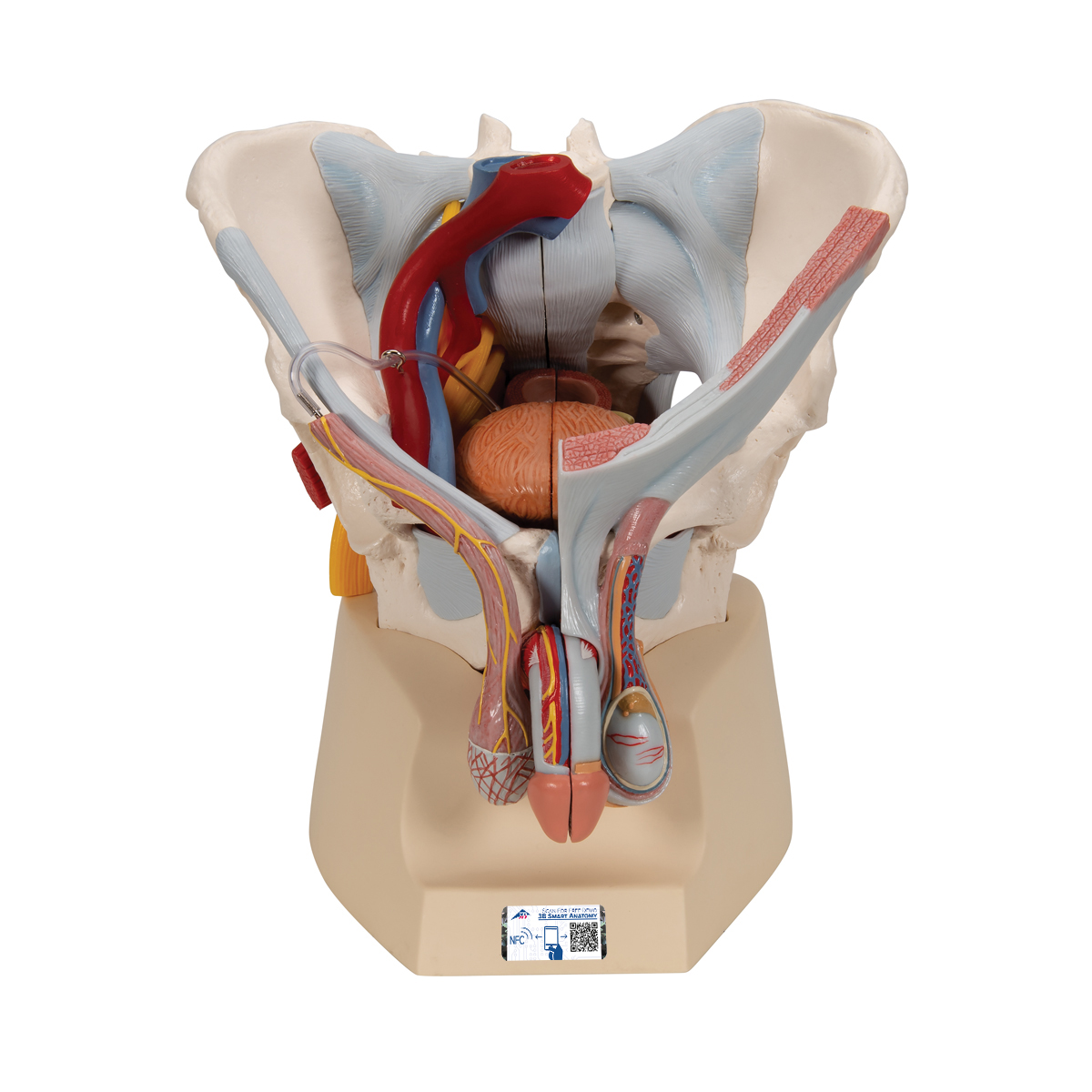
Male Pelvis Skeleton Model With Ligaments Vessels Nerves Pelvic Floor Muscles Organs 7 Part 3b Smart Anatomy 1013282 3b Scientific H21 3 Genital And Pelvis Models Anatomical Models from www.3bscientific.com The pelvic girdle, also known as the hip bone, is composed of three fused bones: The intestines are supported by a series of muscles known as the pelvic. Human anatomy module 4 the pelvis and lower limb. These muscles, including the gluteus maximus and the hamstrings, extend the thigh at the hip in support of the body's weight and propulsion. Just need a glimpse, leave your valuable advice. One is to close the pelvic and abdominal cavities and bear the load of the visceral organs; Arcus tendineus levator ani and the ischial spine: Attached to the pelvis are muscles of the buttocks, the lower back, and the thighs.
The pelvis consists of the sacrum, the coccyx, the ischium, the ilium, and the pubis.
The classification of the two groups under a common heading is. Pelvic floor muscles located wholly within the pelvis One is to close the pelvic and abdominal cavities and bear the load of the visceral organs; Just need a glimpse, leave your valuable advice. The pelvic region holds major organs under its layers of muscles. The pelvic region is the area between the trunk and the lower extremities or. The small intestine is the longest part of the. 3d anatomy tutorial on the pelvic diaphragm from anatomyzone for more videos, 3d models and notes visit: (1) the obturator internus and the piriformis, which are muscles of the lower extremity, and will be described with these (pages 476 and 477); The pelvic girdle (hip girdle) is formed by a single bone, the hip bone or coxal bone (coxal = hip), which serves as the attachment point for each lower limb. (2) the levator ani and the coccygeus, which together form the pelvic diaphragm and are associated with the pelvic viscera. The floor of the pelvis is formed by the two muscles named levator ani and coccygeus. These muscles, including the gluteus maximus and the hamstrings, extend the thigh at the hip in support of the body's weight and propulsion.
Schau dir angebote von muscle anatomy auf ebay an. They form a large sheet of skeletal muscle that is thicker in some areas than in others. 3d anatomy tutorial on the pelvic diaphragm from anatomyzone for more videos, 3d models and notes visit: The floor of the pelvis is formed by the two muscles named levator ani and coccygeus. The ilium, ischium and the pubic bone.
Exploring The Pelvis 3d Muscle Lab from 3dmusclelab.com The classification of the two groups under a common heading is. They form a large sheet of skeletal muscle that is thicker in some areas than in others. Arcus tendineus levator ani and the ischial spine: The muscles of the pelvis and hip control the vast range of movement of the legs and torso. It is usually divided into two separate anatomic regions: Now that you watched the video, you shou. The ilium, ischium and the pubic bone. Of human anatomy and different types of motion, inspiring more realistic and energetic figurative art.
The pelvic region holds major organs under its layers of muscles.
3d anatomy tutorial on the pelvic diaphragm from anatomyzone for more videos, 3d models and notes visit: The term pelvis is used to identify the area between the abdomen and the lower extremities.it can be divided into the greater pelvis and the lesser pelvis. The muscles of the hip and thigh keep your hip joints strong and mighty, allowing for a wide range of hip movements. The pelvic girdle (hip girdle) is formed by a single bone, the hip bone or coxal bone (coxal = hip), which serves as the attachment point for each lower limb. To achieve both these tasks, the pelvic floor is composed of several overlapping sheets of muscles and connective tissues. In this image, you will find glutes medius, superior gemellus, piriformis, gluteus maximus, superior gemellus, obturator internus, ischial spine, attachment to tendon of obturator internus in it. One is to close the pelvic and abdominal cavities and bear the load of the visceral organs; Beginning with the pelvic girdle, the study will move on to the muscles of the pelvis and hip. Just need a glimpse, leave your valuable advice. Along the anterolateral wall of the true pelvis lies the obturator internus muscle. The pelvic girdle, also known as the hip bone, is composed of three fused bones: If you're curious to know more, check out the full. The right and left hip bones also converge anteriorly to attach to each other.
In this image, you will find glutes medius, superior gemellus, piriformis, gluteus maximus, superior gemellus, obturator internus, ischial spine, attachment to tendon of obturator internus in it anatomy muscles pelvis. Attached to the pelvis are muscles of the buttocks, the lower back, and the thighs.


0 Komentar